Radiographs, photographs and endoscopy video — in one viewer.
PrismaVet builds imaging software for veterinary medicine. Every image a patient generates opens in the same browser window, current study and priors together. It runs in the cloud, so there is nothing to install and no server in the building.
Radiographs
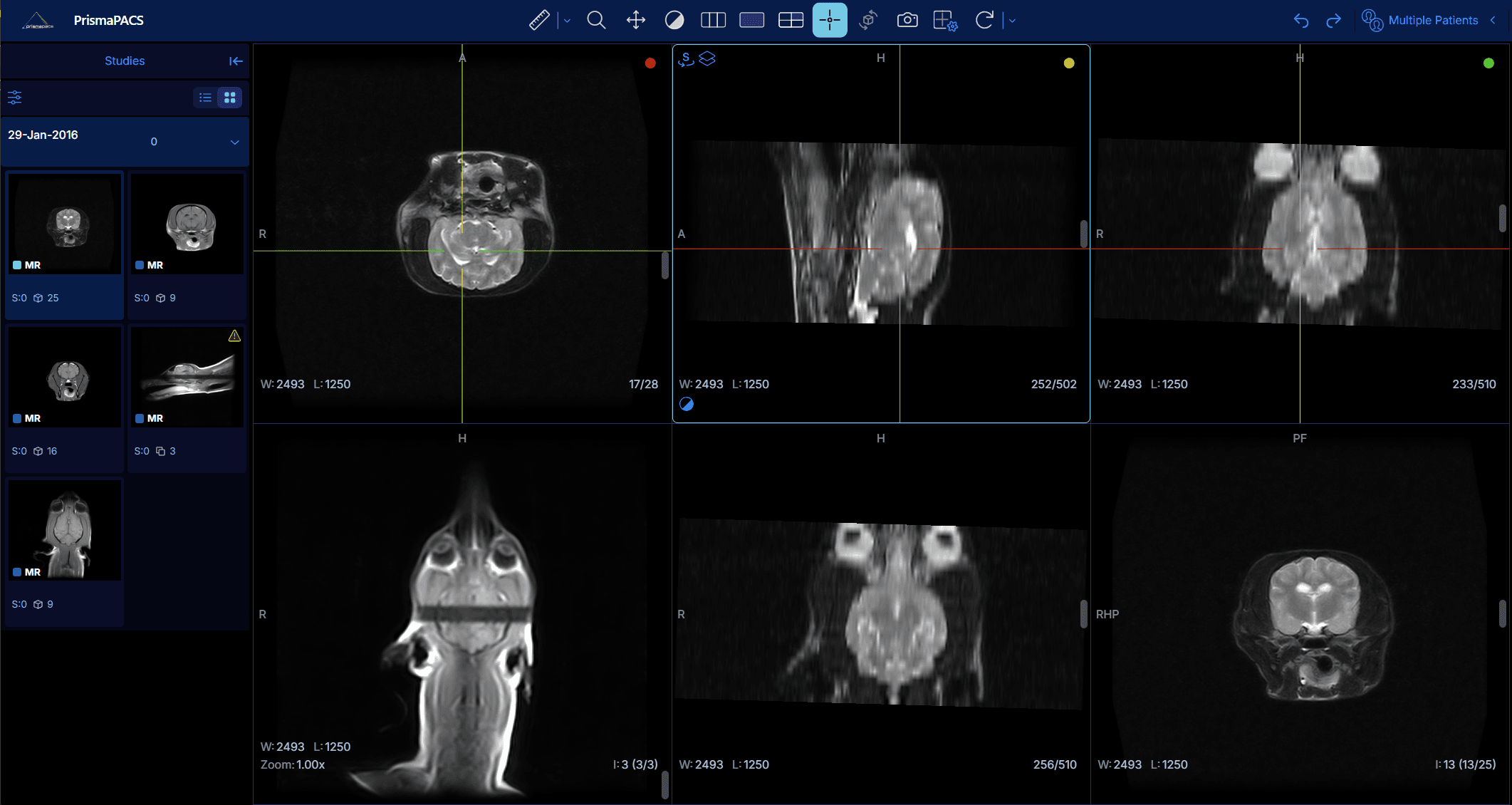
X-ray, CT, ultrasound and MRI, stored at full fidelity and read in the browser — no workstation software and no plug-in.
Visible light
Photographs from dermatology, ophthalmology, surgery and gross pathology live on the patient record, not in a folder on someone's phone.
Endoscopy video
Video studies play in the same viewer as the radiographs, so the whole examination sits in one place instead of two systems.
What PrismaPACS does
01 / CapabilitiesNothing to install, nothing to maintain
PrismaPACS is cloud-based. There is no workstation build, no per-machine license and no server to keep running — connecting a site's imaging equipment takes minutes, and updates arrive on their own.
Priors sit with the current study
Everything a patient has ever had imaged stays on one record. Comparing today's radiograph against last year's is a click, not a search through an archive.
MPR, MIP and 3D, built automatically
Cross-sectional studies are reconstructed as they load. Multiplanar and maximum-intensity views and 3D renderings are ready when the study opens, rather than waiting on someone to reprocess it.
AI assessment in the same window
The viewer integrates with AI-based image assessment, so results appear against the study being read instead of arriving separately and needing to be matched back up.
Studies arrive on their own
A secure connector moves images off the imaging equipment as they are captured, so a study is on the patient record without anyone having to send it there.
Built for groups and referral networks
Run every site from one place, each with its own users and administrators, and keep a record of who opened which study and when.
What EquiLens does
02 / Equine salesA sale repository exists so a horse is radiographed and scoped once, not twenty times. The consignor's veterinarian deposits one set of studies against the hip; every prospective buyer's veterinarian reads that same set before the horse sells. EquiLens is that repository, run by the sale company — across four portals, for staff, submitting vets, buyers' vets and consignors.
Nothing missing at the deadline
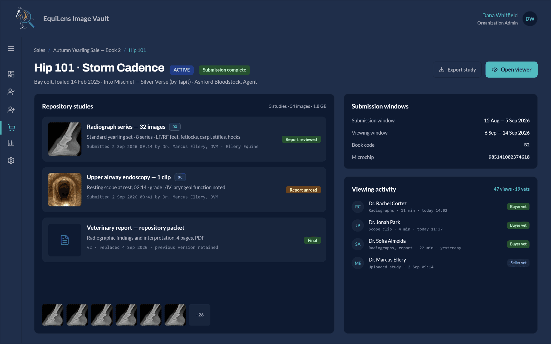
Submitting vets drop a study folder and it is matched to a hip before it lands. Every entry carries its own submission state, so a gap in the repository is visible from the dashboard rather than discovered on sale day.
The whole examination, in one packet
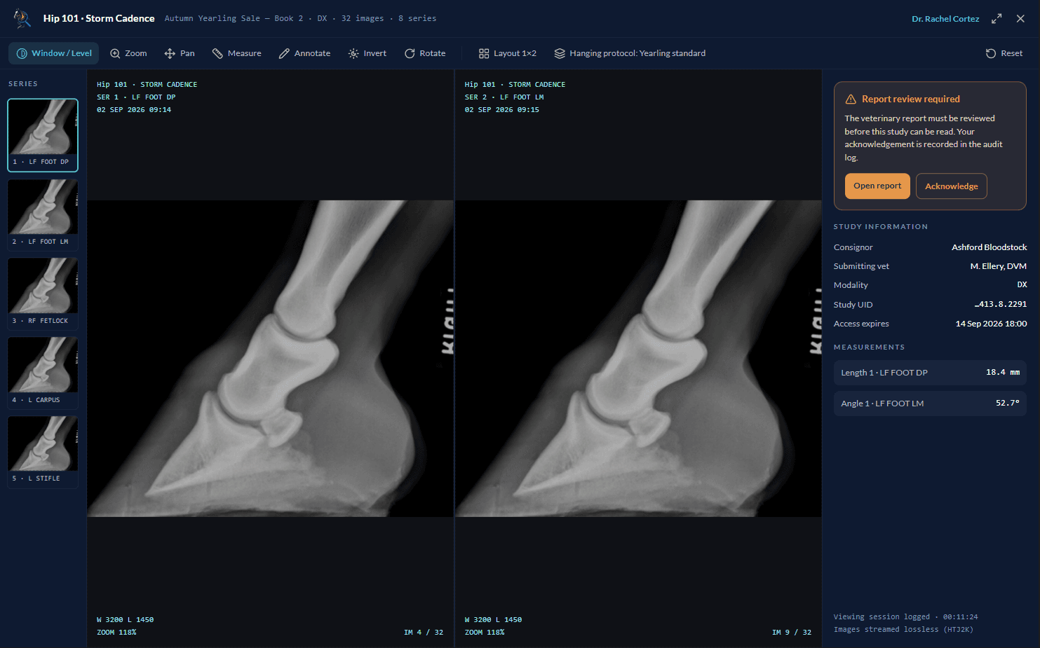
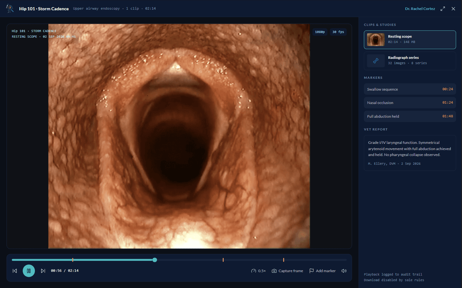
Radiograph series, upper-airway endoscopy and the veterinary report sit together, each with its submitting vet and timestamp. Scope video plays at half speed with markers on the swallow and the abduction, and the vet's grade sits beside the clip rather than in a separate document.
Who read it, and for how long
Every viewing is recorded — vet, series, dwell time, date. Interest in a horse becomes something you can see per hip while the sale is still running, and downloads can be switched off sale by sale.
Where do you fit?
03 / Who it's forPractices & radiology groups
Store, read and share every study your practice produces — across one site or twenty — and give referring vets a link instead of a disc.
Explore PrismaPACS →Equine sale companies
Run the imaging side of a sale: consignor submissions against your deadline, prescribed views checked on arrival, and logged review by buyers' registered veterinarians.
Explore EquiLens →Imaging equipment suppliers
Offer study storage and a browser-based viewer under your own brand, with per-tenant isolation, branded sign-in and site-level administration.
Talk to us →About PrismaVet
04 / AboutPrismaVet builds imaging software for veterinary medicine, and nothing else. Two products share one viewer: PrismaPACS for practices and radiology groups, and EquiLens for equine sale companies.
Veterinary imaging is spread thin — across locations, across modalities, and across vendors who each solve one piece of it. A practice ends up with radiographs in one system, photographs on somebody's phone, scope video on a laptop and priors on a disc in a drawer. We build the part that holds all of it together and puts it in front of whoever has to read it.
The founding team pairs veterinary practice with medical imaging engineering: people who have worked in clinics and read studies, alongside people who have built healthcare imaging systems and the integrations they run on.
Strategic executive and former equine clinic owner, previously COO at a leading veterinary imaging company.
Medical imaging systems engineer, working in healthcare cloud architecture and imaging platform integration.
Medical informatics, healthcare interoperability, and scaling complex system implementations.
Veterinary imaging specialist, focused on AI-assisted diagnostics and digital imaging.
Veterinary technician with 20+ years in digital radiology and software development, and founder of a veterinary consulting firm with specialized equine training.
Your imaging in PrismaPACS
Tell us how images move through your practice today — from the equipment to whoever reads them — and we'll show you that same workflow in PrismaPACS. Connecting the equipment you already run takes minutes, not a migration project.